Fundamentals of Ultrasound

ULS001

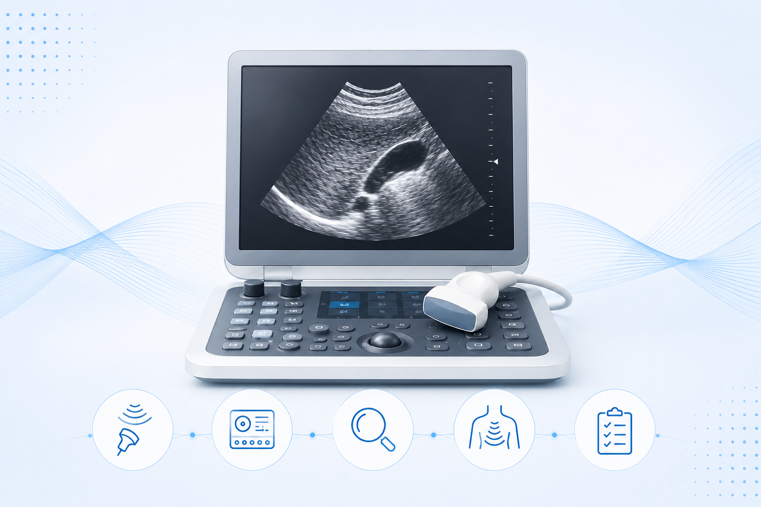
This course provides a comprehensive introduction to the fundamental principles of ultrasound physics and image formation. It covers essential concepts including wave propagation, tissue interactions, transducer technology, image optimization, Doppler ultrasound, harmonic imaging, and common imaging artifacts.

Course Access Period: Course access begins upon purchase, and you will have 30 days to complete the course.


Course Instructors
Dr Abdul Alim Abdul Haium
Dr-Gani-profile-4
Dr-Yasser-Elsayed-profile-1
Dr-Nadya-New
Dr-ASEM-LASHIN-New-1
Overview

About the Course

Master the fundamentals of ultrasound physics, image formation, Doppler principles, and image optimization through practical, interactive learning.

Objectives:

After completing this course, you will be able to:

  • Explain the fundamental principles of ultrasound physics.
  • Apply ultrasound physics principles to optimize image acquisition and Doppler.
  • Integrate ultrasound physics concepts to accurately interpret B-mode and Doppler images.
Course Includes
USD 99.00
Curriculum

Course Content

  • Navigate the AI integrated educational features.
  • Identify and understand the functions of the simulator interface and ultrasound machine controls (knobology).
  • Use the simulator confidently to support ultrasound learning and image optimization.
  • Explain the basic principles of ultrasound physics
  • Describe the relationship between ultrasound frequency, penetration, and spatial resolution
  • Explain the function of piezoelectric crystals
  • Describe the process of pulse transmission, echo reception, and image formation
  • Interpret how tissue properties influence echo generation and grayscale image appearance
  • Explain the fundamental physics of ultrasound image formation.
  • Optimize ultrasound image quality.
  • Describe the principles of Doppler ultrasound.
  • Recognize and interpret common ultrasound artifacts.
  • Comprehensive video lesson on Ultrasound Physics and the visual language of sonography.
  • Explore knobology, doppler, artifacts, image interpretation.
  • Explain the fundamental physical properties of ultrasound waves.
  • Differentiate operator-controlled parameters from tissue-dependent properties .
  • Apply the principles of ultrasound physics to clinical imaging.
  • Explain the principles of pulse-echo ultrasound.
  • Differentiate the key pulse-echo parameters.
  • Apply the relationship between imaging depth and pulse timing.
  • Explain the physical principles governing sound wave propagation .
  • Analyze the interdependence of frequency, wavelength, and speed of sound.
  • Apply the principles of sound-wave physics.
  • Explain the relationships between pressure, power, and ultrasound intensity.
  • Describe the effects of attenuation and the decibel scale.
  • Interpret spatial and temporal intensity measurements.
  • Define acoustic impedance (Z) .
  • Explain the three primary interactions of ultrasound with tissue boundaries.
  • Describe how acoustic impedance differences (ΔZ) determine echo production.
  • Identify the factors that determine acoustic impedance.
  • Explain how acoustic impedance mismatch determines reflection and transmission.
  • Calculate reflected and transmitted ultrasound energy using the reflection coefficient.
  • Explain why beam angle affects echo detection and image quality.
  • Differentiate the mechanisms of acoustic shadowing caused by air and bone.
  • Define attenuation and distinguish the ultrasound properties that decrease with tissue travel from those that remain constant.
  • Explain the mechanisms of scatter and absorption and their roles in attenuation and image formation.
  • Describe the factors affecting attenuation, including depth, frequency, and tissue type.
  • Define axial resolution and explain its importance in distinguishing structures at different depths.
  • Explain how dampening improves axial resolution by reducing the number of cycles per pulse.
  • Identify the factors that determine axial resolution and explain why it remains constant with imaging depth.
  • Differentiate A-mode, B-mode, and M-mode based on their display format, dimensions, and clinical applications.
  • Explain how ultrasound echoes are converted into diagnostic images.
  • Describe how echo amplitude and time-of-flight determine image brightness and depth.
  • Differentiate lateral and elevational resolution and describe their roles in resolving structures at the same depth.
  • Explain how beam width and beam height determine lateral and elevational resolution, respectively.
  • Describe the effects of focusing and beam geometry on image resolution, including depth dependence.
  • Describe the structure and function of modern PZT transducer arrays.
  • Explain the differences between linear sequential arrays and phased arrays.
  • Describe how electronic beam steering is achieved in phased-array transducers.
  • Explain the fundamental physics of ultrasound.
  • Apply the principles of ultrasound wave behavior.
  • Differentiate the major ultrasound imaging modes, probe types, and transducer technologies.
  • Explain the fundamental principles of ultrasound physics.
  • Apply the physical principles of ultrasound to optimize image acquisition.
  • Interpret ultrasound images and Doppler data by integrating knowledge of tissue interactions.
  • Apply ultrasound physics to optimize image acquisition.
  • Interpret ultrasound images and Doppler findings.
  • Explain the Doppler effect and Doppler shift and their relationship to blood flow velocity.
  • Apply the Doppler equation to describe how transducer frequency, blood velocity, and insonation angle influence Doppler measurements.
  • Identify and interpret the components of a spectral Doppler waveform.
  • Explain how PRF, gate size, Doppler angle, and the Nyquist limit.
  • Explain the principles of Tissue Harmonic Imaging (THI).
  • Describe how harmonic signal isolation improves image quality.
  • Compare conventional pulse-echo imaging with Tissue Harmonic Imaging.
  • Explain the physical assumptions underlying ultrasound image formation.
  • Identify and differentiate common ultrasound artifacts.
  • Describe the physical mechanisms responsible for each artifact.
  • Recall and define the fundamental concepts, terminology, and equations of ultrasound physics.
  • Apply ultrasound physics principles to solve clinical and image interpretation problems.
  • Navigate the ultrasound simulator confidently, including orientation, workflow, and system interface for efficient image acquisition.
  • Demonstrate proficiency in ultrasound knobology by identifying and appropriately adjusting all major machine controls
Faculty

Course Instructors

Prof. Yasser Elsayed

Prof. Yasser Elsayed

Professor
University of Manitoba
Winnipeg, Canada
Similar

Other Courses

Master the fundamentals of neonatal lung ultrasound, including ultrasound language, scanning techniques, lung ultrasound

USD 99.00

Master foundational neonatal hemodynamics, echocardiography, blood pressure assessment, and physiology through practical interactive learning.

USD 99.00

Build confidence assessing neonatal hemodynamics and managing complex cardiovascular conditions through advanced echocardiographic skills.

USD 99.00

Master pulmonary hypertension physiology, phenotypes, diagnosis, management, and non-invasive treatment across neonates and children.

USD 99.00
x